Volume: 3 Issue: 2
Antenatal Pleural Effusion: Diagnostic Challenges and Management as Presumptive Tubercular Disease
Year: 2026, Page: 71-73, Doi: https://doi.org/10.71325/ajjms.v3i2.26.27
Received: June 25, 2026 Accepted: Aug. 6, 2026 Published: Aug. 12, 2026
Abstract
Background: Tuberculosis in pregnancy remains a diagnostic challenge, especially in extrapulmonary forms. Pleural effusion with lymphocyte predominance often raises suspicion for tubercular etiology, even when microbiological tests are negative. Case Presentation: A 38 year old G2P1L1 woman at 29 weeks and 6 days of gestation who presented with fever, excessive vomiting, and generalized weakness. There were no initial respiratory complaints. Routine obstetric ultrasound incidentally revealed pleural effusion. Further evaluation was undertaken, and diagnostic pleural tapping was performed. Pleural fluid analysis showed lymphocyte-predominant exudative effusion. Cytology, CBNAAT, and culture sensitivity were negative for infectious organisms, including Mycobacterium tuberculosis. Management and Outcome: In view of persistent symptoms and lymphocytic predominance of pleural fluid, a presumptive diagnosis of tubercular pleural effusion was made. The patient was started on weight-based anti-tubercular therapy (ATT). She showed clinical improvement with reduction in systemic symptoms on follow-up. Pregnancy was continued with close maternal and fetal monitoring. Conclusion: A high index of suspicion is essential for diagnosing tuberculosis in pregnancy, even in the absence of microbiological confirmation. Empirical ATT based on clinical and biochemical findings can lead to favorable outcomes when initiated timely.
Keywords: Extrapulmonary TB in pregnancy, Microbiologically negative, Empirical ATT
INTRODUCTION
Tuberculosis (TB) remains a major cause of maternal morbidity in regions where the disease is endemic[1]. Pregnancy-associated physiological and immunological changes may modify the clinical presentation of TB, often resulting in delayed diagnosis or under recognition[1]. Extrapulmonary tuberculosis, particularly tuberculous pleural effusion, presents a significant diagnostic challenge because of its nonspecific clinical features and the limited sensitivity of conventional microbiological investigations, including smear microscopy, mycobacterial culture, and cartridge-based nucleic acid amplification testing (CBNAAT)[2, 3]. In such cases, the presence of an exudative, lymphocyte-predominant pleural effusion strongly suggests a tuberculous etiology, even when microbiological confirmation is unavailable or inconclusive[4, 5].
CASE REPORT
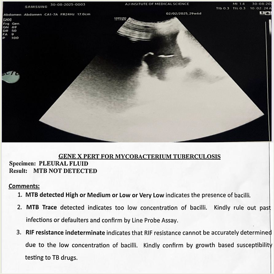
A 38-year-old G2P1L1 woman at 29 weeks and 6 days of gestation presented with complaints of fever, excessive vomiting, and generalized weakness for one week. There were no associated respiratory symptoms such as cough, breathlessness, or chest pain. Her antenatal period had been uneventful prior to this presentation. On examination, the patient was hemodynamically stable. Systemic examination revealed reduced breath sounds over left Infra axillary area / Infra Scapular Area / Infra Mammary Area. Obstetric examination corresponded to the period of gestation, and fetal heart rate was within normal limits. Routine obstetric ultrasonography incidentally detected pleural effusion, prompting further evaluation. USG of Bilateral Hemithorax revealed left side pleural effusion of 2.5-2.7 litres with collapse of left lower lobe. Diagnostic thoracentesis was performed. Pleural fluid analysis revealed an exudative effusion with lymphocyte predominance. Cytological examination was negative for malignant cells. Microbiological investigations, including smear microscopy, culture sensitivity, CBNAAT and GeneXpert were negative for infectious organisms, including Mycobacterium tuberculosis. In view of raised Adenosine deaminase (ADA – 70 U/L) and lymphocyte-rich exudative pleural effusion, a presumptive diagnosis of tubercular pleural effusion was made. The patient was initiated on weight- based anti-tubercular therapy (ATT). Following initiation of therapy, the patient showed significant clinical improvement with reduction in systemic symptoms. She was maintained on regular antenatal follow-up with close maternal and fetal monitoring, and the pregnancy was continued. At term scan EFW was <5th centile indicating SGA. Interval growth scans were done and fetal well being assessed. Elective LSCS in view of previous LSCS at 38 weeks. Postnatally, baby was started on Isoniazid 25mg OD prophylaxis.
DISCUSSION
Tuberculosis (TB) during pregnancy remains a significant diagnostic challenge, particularly in cases of extrapulmonary involvement where symptoms are often nonspecific[1, 2]. Pleural tuberculosis is one of the most common forms of extrapulmonary TB and typically presents with fever, constitutional symptoms, and pleural effusion[4, 5]. However, in pregnancy, classical respiratory symptoms may be absent or masked, as seen in our case[1]. Our patient presented with fever, vomiting, and generalized weakness without respiratory complaints. The diagnosis was incidentally suspected following obstetric ultrasonography, which revealed pleural effusion. This highlights the importance of maintaining a high index of suspicion for TB in endemic regions like India, even when symptoms are atypical[1, 5].
Pleural fluid analysis in tuberculous pleural effusion classically demonstrates a lymphocyte-predominant exudate with elevated protein levels[4, 5]. However, microbiological confirmation using CBNAAT, culture, or smear microscopy is frequently negative because of the paucibacillary nature of the disease[2, 4]. In such cases, the diagnosis is based on a combination of clinical suspicion, pleural fluid biochemical findings, and supportive radiological features.2,4 Our case demonstrated a lymphocyte-rich exudative pleural effusion with negative microbiological investigations, consistent with findings reported in previous studies[4, 5].
Empirical initiation of anti-tubercular therapy (ATT) in patients with a high clinical suspicion is recommended by national and international guidelines, particularly when alternative diagnoses have been reasonably excluded[1, 2, 6]. The favorable clinical response to ATT in our patient further supports the diagnosis. First-line anti-tubercular drugs are considered relatively safe during pregnancy and are associated with favorable maternal and fetal outcomes when treatment is initiated promptly[1, 6].
CONCLUSION
Extrapulmonary tuberculosis in pregnancy can present with non-specific and atypical symptoms, often without classic respiratory features. This case highlights the diagnostic challenge posed by microbiologically negative pleural effusion, where a high index of clinical suspicion is crucial. Lymphocyte-predominant exudative effusion should prompt consideration of tuberculosis, especially in endemic regions. Early initiation of empirical anti-tubercular therapy in such cases can lead to favourable maternal and fetal outcomes. Timely diagnosis and management are essential to reduce morbidity and improve prognosis.
DISCLOSURE
Conflict of Interest: None.
Funding: Nil.
References
1. World Health Organization. WHO recommendations on antenatal care for a positive pregnancy experience: screening, diagnosis and treatment of tuberculosis disease in pregnant women. Geneva: World Health Organization; 2023.
2. World Health Organization. WHO consolidated guidelines on tuberculosis. Module 3: Diagnosis. Geneva: World Health Organization; 2025.
3. Lewinsohn DM, Leonard MK, LoBue PA, Cohn DL, Daley CL, Desmond E, et al. Official American Thoracic Society/Infectious Diseases Society of America/Centers for Disease Control and Prevention Clinical Practice Guidelines: Diagnosis of Tuberculosis in Adults and Children. Clinical Infectious Diseases. 2017; 64 (2). Available from: https://doi.org/10.1093/cid/ciw778
4. Light RW. Light's Pleural Diseases. 7th ed. Philadelphia: Wolters Kluwer; 2022.
5. Sharma SK, Mohan A. Extrapulmonary tuberculosis. Indian Journal of Medical Research. 2004;120(4):316-53.
6. Ministry of Health and Family Welfare, Government of India. National Tuberculosis Elimination Programme (NTEP): Technical and Operational Guidelines. New Delhi: Central TB Division; 2021.
Copyright
© 2026 Published by Laxmi Memorial Education Trust. This is an open-access article under CC BY 4.0 license. (https://creativecommons.org/licenses/by/4.0/)
Cite this article
Dhanya Uday Nayak, Anitha S. Antenatal Pleural Effusion: Diagnostic Challenges and Management as Presumptive Tubercular Disease. AJ J Med Sci 2026;3(2):71-73