Volume: 3 Issue: 2
Insights into Virulence and Pathogenesis of Staphylococcus aureus and Mycobacterium tuberculosis: A Comprehensive Review
Year: 2026, Page: 50-56, Doi: https://doi.org/10.71325/ajjms.v3i2.26.31
Received: July 13, 2026 Accepted: Aug. 6, 2026 Published: Aug. 12, 2026
Abstract
Bacterial pathogens employ diverse virulence factors to establish infection, evade host defences and promote disease progression. This review examines the virulence strategies of two clinically significant human pathogens, Staphylococcus aureus and Mycobacterium tuberculosis, which represent contrasting paradigms of bacterial infection. S. aureus utilizes an extensive repertoire of adhesins, exotoxins, immune evasion molecules and biofilm-forming mechanisms to induce rapid tissue destruction and systemic disease. In contrast, M. tuberculosis relies on complex cell wall components, intracellular survival mechanisms, immune modulation and dormancy associated pathways to establish persistent infection. Key virulence determinants include Microbial Surface Components Recognizing Adhesive Matrix Molecules (MSCRAMMs), protein A, α-toxins, quorum sensing systems, mycolic acids, lipoarabinomannan, ESX secretion systems and the DosR regulon. A comparative analysis highlights fundamental differences in acute and chronic infection strategies adopted by these pathogens. Understanding these distinct virulence strategies may facilitate the development of novel anti-virulence therapies and host-directed interventions, particularly in the context of rising antimicrobial-resistant pathogens.
Keywords: Virulence factors, Staphylococcus aureus, Mycobacterium tuberculosis, Pathogenesis, Immune evasion, Biofilms, Anti-virulence therapy, Toxins, Intracellular survival
INTRODUCTION
Bacterial virulence factors comprise specialized structural components, secreted effectors, enzymes, and regulatory networks that enable pathogens to colonize host tissues, evade immune responses, and establish disease[1]. These factors coordinate critical stages of infection, including adhesion, invasion, immune modulation and dissemination.
Among bacterial pathogens, Staphylococcus aureus and Mycobacterium tuberculosis represent two contrasting yet globally important models of infection. Staphylococcus aureus is a Gram-positive opportunistic pathogen responsible for a wide spectrum of diseases ranging from superficial skin and soft tissue infections to life threatening endocarditis, osteomyelitis, necrotizing pneumonia and sepsis[2]. Its success as a pathogen is attributed to its diverse range of virulence factors and its ability to acquire antimicrobial resistance, exemplified by Methicillin-resistant S. aureus (MRSA). Recent studies have demonstrated that S. aureus primarily relies on adhesins, exotoxins, immune evasion molecules and biofilm formation, contributing to persistence, recurrence and treatment failure[3].
Conversely, Mycobacterium tuberculosis, the etiological agent of tuberculosis, one of the leading infectious causes of mortality worldwide is a slow-growing intracellular pathogen that primarily infects macrophages. M. tuberculosis’s virulence stems from its ability to survive inside host cells through a lipid-rich cell envelope, manipulate ESX secretion systems, form granulomas and establish dormancy[4, 5].
The selection of Staphylococcus aureus and Mycobacterium tuberculosis for comparison is particularly relevant because they represent two fundamentally different strategies of bacterial pathogenesis. While S. aureus causes predominantly acute, toxin-mediated infections, M. tuberculosis establishes chronic intracellular infection through immune modulation and persistence. Both organisms remain major contributors to global morbidity and antimicrobial resistance, making them important models for studying virulence evolution and therapeutic innovation[6].
This review provides valuable insights into conserved and divergent virulence mechanisms and highlights opportunities for anti-virulence and host-directed therapeutic interventions.
Virulence Factors and Pathogenesis of Staphylococcus aureus
Adhesion and Colonization
Successful infection by S. aureus begins with adhesion to host tissues. This process is mediated by a family of surface proteins called Microbial Surface Components Recognizing Adhesive Matrix Molecules (MSCRAMMs)[7]. Important members include fibronectin-binding proteins (FnBPA and FnBPB), clumping factors (ClfA and ClfB) and the collagen-binding protein (Cna).
These proteins facilitate attachment to extracellular matrix components such as fibronectin, fibrinogen and collagen, promoting colonization and invasion of host tissues. ClfA, for example, binds fibrinogen and plays a crucial role in bloodstream infections by enhancing bacteria aggregation[8].
Immune Evasion Mechanism
Following colonization, S. aureus employs sophisticated tactics to evade host immune responses and contribute to chronic infections, emphasizing that S. aureus should no longer be considered as an exclusively extracellular pathogen[9, 10]. These tactics include interfering with complement activation, neutralizing antibodies, and manipulating host cell signaling pathways.
Protein A binds to the Fc region of immunoglobulin G (IgG), thereby impairing opsonization and phagocytosis[11]. This mechanism effectively disguises bacteria from immune recognition.
Additional immune evasion factors include the staphylococcal complement inhibitor (SCIN), which inhibits complement activation and the chemotaxis inhibitory protein of staphylococci (CHIPS) which disrupts neutrophil recruitment. Together, these factors reduce the effectiveness of innate immune responses and facilitate bacterial survival[12].
Exotoxins and Tissue Damage
A hallmark of S. aureus pathogenesis is the production of potent exotoxins that directly damage host tissues. Alpha-toxin (α-hemolysin) forms pores in the membranes of host cells, resulting in cell lysis and tissue destruction. This toxin is particularly important in pneumonia and skin infections.
Panton-Valentine leukocidin (PVL) specifically targets leukocytes, promoting immune cell destruction and enhancing bacterial survival. Toxic shock syndrome toxin-1 (TSST-1) and enterotoxins function as superantigens, triggering non-specific T-cell activation and massive cytokine release leading to systemic inflammation.
Emerging research suggests that toxin-mediated virulence is often synergistic rather than isolated. Interactions between α-toxins, PVL and other cytotoxins amplify tissue injury and disease severity, particularly in highly virulent MRSA strains[13].
Enzymatic Virulence Factors
S. aureus secretes several enzymes that aid in tissue invasion and nutrient acquisition. Coagulase promotes fibrin clot formation, providing a protective niche around bacterial colonies. Hyaluronidase degrades connective tissue matrices, while proteases and lipases digest host macromolecules[14].
Biofilm formation
Biofilm formation represents a major virulence strategy that contributes to chronic infection and antibiotic tolerance by reducing antibiotic penetration and enhancing bacterial persistence. This process begins with adhesion mediated by MSCRAMMs and progresses through bacterial aggregation and accumulation of polysaccharide intercellular adhesin (PIA)[15, 16]. Biofilms-associated infections are particularly problematic in indwelling medical devices such as catheters, prosthetic joints and cardiac implants.
Emerging evidence suggests that biofilm architecture is highly dynamic and regulated by environmental stress, quorum sensing, and metabolic adaptation, contributing significantly to antibiotic tolerance and immune evasion.
Regulation of Virulence
The expression of virulence determinants is tightly controlled through global regulatory systems. The accessory gene regulator (agr) system is a quorum-sensing mechanism that regulates toxin production and biofilm formation[17]. Other regulators, including SarA and sigma factor B (σB), modulate stress responses and coordinate the expression of virulence associated-genes.
| Virulence factor | Function | Clinical significance |
|---|---|---|
| MSCRAMMs | Adhesion to extracellular matrix | Colonization |
| Protein A | Fc-binding protein | Immune evasion |
| SCIN | Complement inhibition | Reduced opsonization |
| CHIPS | Inhibits neutrophil chemotaxis | Immune escape |
| α-toxin | Pore-forming toxin | Tissue destruction |
| PVL | Leukocyte destruction | Necrotizing infections |
| TSST-1 | Superantigen | Toxic shock syndrome |
| Coagulase | Fibrin clot formation | Protection from phagocytosis |
| Hyaluronidase | Tissue invasion | Spread of infection |
| agr system | Quorum sensing | Virulence regulation |
| Biofilm (PIA) | Persistence | Device-associated infections |
Table 1: Major virulence factors of Staphylococcus aureus
Virulence Factors and Pathogenesis of Mycobacterium tuberculosis
Cell Wall Components
The unique lipid rich cell wall of M. tuberculosis is a major virulence determinant. Mycolic acid confers resistance to desiccation, disinfectants and many antimicrobial agents.
Lipoarabinomannan (LAM) modulates host immune responses by inhibiting macrophage activation and cytokine production. Another critical lipid, trehalose dimycolate (cord factor), contributes to granuloma formation and tissue pathology[18-20].
These cell wall components not only provide structural protection but also actively participate in host-pathogen interactions.
Intracellular Survival
Unlike S. aureus, M. tuberculosis primarily survives within macrophages. After phagocytosis, the pathogen prevents phagosome-lysosome fusion, thereby escaping intracellular destruction.
The bacteria further resists oxidative and nitrosative stress while manipulating host signaling pathways to create a favorable intracellular environment. Recent studies have demonstrated that M. tuberculosis can alter host cell metabolism and inhibits autophagy, enhancing long-term intracellular persistence[21, 22].
ESX-1 Secretion System
The ESX-1 (Type VII) secretion system is among the most important virulence mechanisms for M. tuberculosis. It secretes proteins such as ESAT-6, which disrupts phagosomal membranes and CFP-10, which stabilizes ESAT-6 and boosts virulence[23].
Beyond membrane disruption, ESX-1 also modulates host immune signaling and contributes to bacterial spread between cells. Studies have shown that mutations affecting ESX-1 components markedly reduce virulence, highlighting its potential as a therapeutic target.
Granuloma Formation and Immune Modulation
Granulomas are structured immune clusters that develop in response to persistent infection. While granulomas restrict bacterial dissemination, they simultaneously provide a protected niche that facilitates long-term persistence.
M. tuberculosis manipulates granuloma dynamics by suppressing antigen presentation, promoting anti-inflammatory cytokines such as IL-10 and inhibiting programmed cell death[24]. Through these mechanisms, the pathogen maintains a delicate balance between immune activation and immune suppression, thereby ensuring long term survival within the host.
Dormancy and Latency
A defining characteristic of M. tuberculosis is its ability to enter a dormant state during latent infection.
The DosR regulon becomes activated under hypoxic and stress conditions, inducing a transcriptional program that promotes long term bacterial survival[25]. Dormant bacilli exhibit reduced metabolic activity and increased resistance to both antimicrobial therapy and immune-mediated clearance. Recent investigations into the DosR regulon and resuscitation-promoting factors have improved understanding of latent tuberculosis and identified potential therapeutic targets for preventing disease reactivation[26].
This ability to establish latency distinguishes M. tuberculosis from many other bacterial pathogens and represents a major challenge for tuberculosis control efforts worldwide. Additionally, this global burden is exacerbated by the rise of multidrug-resistant (MDR-TB) and extensively drug-resistant tuberculosis (XDR-TB).
| Virulence factor | Function | Clinical significance |
|---|---|---|
| Mycolic acids | Cell wall integrity | Drug resistance |
| Lipoarabinomannan (LAM) | Immune modulation | Macrophage dysfunction |
| Cord factor | Granuloma formation | Tissue damage |
| ESX-1 secretion system | ESAT-6 secretion | Intracellular survival |
| CFP-10 | ESAT-6 stabilization | Virulence |
| DosR regulon | Dormancy | Latent TB |
| Antioxidant enzymes | Oxidative stress resistance | Intracellular persistence |
| Lipid metabolism | Nutrient acquisition | Chronic infection |
Table 2: Major virulence factors of Mycobacterium tuberculosis
Emerging Anti-virulence and Host-directed Therapeutic Strategies
Recent therapeutic strategies increasingly target bacterial virulence rather than viability, thereby reducing selective pressure for antimicrobial resistance. For S. aureus, inhibition of the agr quorum-sensing system, neutralization of α-toxin with monoclonal antibodies, anti-biofilm peptides, bacteriophage therapy and vaccines targeting surface adhesins have demonstrated promising preclinical or early clinical results[27, 28]. Similarly, host-directed therapies for tuberculosis seek to augment host immunity instead of directly targeting the bacillus. Agents such as metformin, vitamin D, statins and autophagy-inducing drugs improve macrophage antimicrobial activity and may enhance treatment outcomes[29-32]. Novel inhibitors targeting the ESX-1 secretion system and bacterial dormancy pathways are also under investigation[33, 34]. Integration of host-directed therapies with conventional antimicrobial regimens may shorten treatment duration and reduce emergence of resistance.
Future research should focus on integrating genomic, transcriptomic, and proteomic approaches to better understand host-pathogen interactions and identify novel therapeutic targets.
| Pathogen | Current targets | Emerging targets |
|---|---|---|
| S. aureus | Antibiotics | agr inhibitors, anti-α toxin antibodies, anti-biofilm peptides, bacteriophage therapy, vaccines |
| M. tuberculosis | Standard anti-TB drugs | Host-directed therapy, ESX-1 inhibitors, autophagy enhancers, metformin, statins, vitamin D, immune checkpoint modulation |
Table 3: Emerging therapeutic targets
| Feature | Staphylococcus aureus | Mycobacterium tuberculosis |
|---|---|---|
| Gram stain | Gram-positive coccus | Acid-fast bacillus |
| Primary lifestyle | Extracellular (facultative intracellular) | Obligate intracellular pathogen |
| Reservoir | Skin and nasal mucosa | Human lungs |
| Disease pattern | Acute | Chronic |
| Major virulence factors | MSCRAMMs, Protein A, α-toxin, PVL | Mycolic acids, LAM, ESX-1, DosR |
| Immune evasion | Protein A, SCIN, CHIPS | Phagosome maturation arrest, immune modulation |
| Persistence mechanism | Biofilm, intracellular survival | Granuloma formation, latency |
| Tissue damage | Toxin-mediated | Immune-mediated |
| Drug resistance | MRSA | MDR-TB, XDR-TB |
| Emerging therapies | Anti-toxin antibodies, agr inhibitors | Host-directed therapy, ESX-1 inhibitors |
| Vaccine status | No licensed vaccine | BCG available; newer candidates under evaluation |
| Clinical challenge | Recurrent device-associated infections | Long treatment duration and latent infection |
Table 4: Comparative overview of S. aureus Vs M. tuberculosis
Schematic representation of Staphylococcus aureus pathogenesis
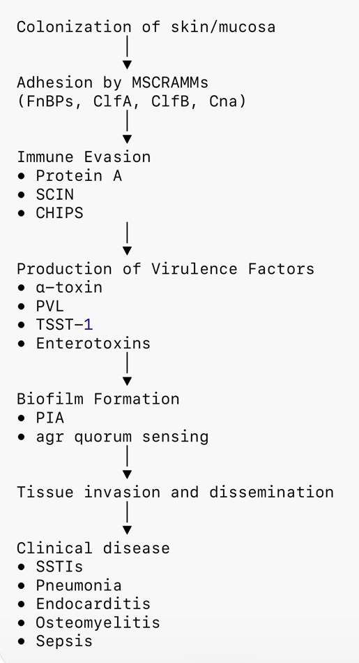
Fig. 1: Major virulence mechanisms of Staphylococcus aureus. Infection begins with adhesion mediated by MSCRAMMs, followed by immune evasion through Protein A and complement inhibitors. Production of cytotoxins and biofilm formation facilitate tissue destruction, persistence, and dissemination, resulting in both localized and invasive infections
Schematic representation of Mycobacterium tuber- culosis pathogenesis
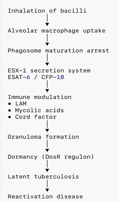
Fig. 2: Major pathogenic mechanisms of Mycobacterium tuberculosis. The organism survives within macrophages by inhibiting phagosome maturation, manipulating host immunity through lipid-rich cell wall components and the ESX-1 secretion system, eventually establishing granulomas and latent infection
DISCUSSION
The two pathogens illustrate distinct evolutionary solutions to host survival. S. aureus adopts an aggressive strategy characterized by rapid adhesion, toxin production and tissue destruction that promotes dissemination before adaptive immunity is fully established. In contrast, M. tuberculosis prioritizes immune modulation, intracellular survival and metabolic adaptation, enabling persistence for decades[35]. These divergent lifestyles explain why therapies that neutralize toxins or disrupt biofilms are particularly attractive for S. aureus, whereas tuberculosis management requires enhancement of host immunity and disruption of bacterial persistence.
Despite these differences, both pathogens manipulate innate immunity, establish protected niches and display remarkable adaptability under antimicrobial pressure. Increasing antimicrobial resistance, including MRSA and multidrug-resistant tuberculosis, highlights the limitations of conventional antibiotics. Consequently, anti-virulence and host-directed therapeutic strategies have emerged as complementary approaches. Several innovative strategies are currently under investigation, including monoclonal antibodies against α-toxin, agr quorum-sensing inhibitors, bacteriophage therapy and anti-biofilm compounds for S. aureus. For M. tuberculosis, host-directed therapies involving metformin, statins, vitamin D supplementation, autophagy modulation and immune checkpoint regulation have demonstrated encouraging preclinical and early clinical outcomes.
However, translation into clinical practice remains limited because bacterial virulence is multifactorial and varies among strains. Furthermore, advances in genomics, transcriptomics, proteomics and artificial intelligence-assisted drug discovery are expected to accelerate identification of novel virulence determinants and facilitate precision antimicrobial development[36]. Comparative analyses such as this review emphasize that successful antimicrobial strategies must account for pathogen-specific biology rather than relying on a single universal approach.
Collectively, these observations underscore that future antimicrobial strategies should move beyond bactericidal activity alone and increasingly target virulence regulation, host–pathogen interactions, and bacterial persistence.
CONCLUSION
Staphylococcus aureus and Mycobacterium tuberculosis represent contrasting yet highly successful models of bacterial pathogenesis. While S. aureus relies on adhesion, toxin-mediated tissue destruction, and biofilm formation to establish acute infections, M. tuberculosis achieves intracellular persistence through immune modulation, granuloma formation and dormancy. Comparative evaluation of these pathogens demonstrates that distinct virulence strategies require equally distinct therapeutic approaches. Recent advances in anti-virulence therapy, host-directed interventions and precision antimicrobial development provide promising alternatives to conventional antibiotics. Continued multidisciplinary research integrating molecular microbiology, immunology and translational medicine will be essential for combating antimicrobial resistance and improving clinical outcomes.
References
1. Finlay BB, Falkow S. Common themes in microbial pathogenicity revisited. Microbiology and Molecular Biology Reviews. 1997; 61 (2). Available from: https://doi.org/10.1128/mmbr.61.2.136-169.1997
2. Lowy FD. Staphylococcus aureus infections. New England Journal of Medicine. 1998; 339 (8). Available from: https://doi.org/10.1056/nejm199808203390806
3. Rodrigues Lopes I, Alcantara LM, Lopez-Bravo M. et al. Systematic identification of bacterial factors driving Staphylococcus aureus intracellular lifestyle in non-professional phagocytes. Nature Communications. 2025; 16 (1). Available from: https://doi.org/10.1038/s41467-025-66373-9
4. Rahlwes KC, Shiloh MU. Pathogenicity and virulence of Mycobacterium tuberculosis. Virulence. 2023; 14 (1). Available from: https://doi.org/10.1080/21505594.2022.2150449
5. Sun MR, Xing JY, Li XT, Fang R, Zhang Y, Li ZL, et al. Recent advances in research on Mycobacterium tuberculosis virulence factors and their role in pathogenesis. Journal of Microbiology, Immunology and Infection. 2025; 58 (5). Available from: https://doi.org/10.1016/j.jmii.2025.03.017
6. Clatworthy AE, Pierson E, Hung DT. Targeting virulence: a new paradigm for antimicrobial therapy. Nature Chemical Biology. 2007; 3 (9). Available from: https://doi.org/10.1038/nchembio.2007.24
7. Foster TJ, Hook M. Surface protein adhesins of Staphylococcus aureus. Trends in Microbiology. 1998; 6 (12). Available from: https://doi.org/10.1016/s0966-842x(98)01400-0
8. McDevitt D, Francois P, Vaudaux P, Foster TJ. Molecular characterization of the clumping factor (fibrinogen receptor) of Staphylococcus aureus. Molecular Microbiology. 1994; 11 (2). Available from: https://doi.org/10.1111/j.1365-2958.1994.tb00304.x
9. Rooijakkers SHM, van Kessel KPM, van Strijp JAG. Staphylococcal innate immune evasion. Nature Reviews Microbiology. 2005;3(12):948–958.
10. Cheung GYC, Bae JS, Otto M. Pathogenicity and virulence of Staphylococcus aureus. Virulence. 2021; 12 (1). Available from: https://doi.org/10.1080/21505594.2021.1878688
11. Palmqvist N, Foster T, Tarkowski A, Josefsson E. Protein A is a virulence factor in Staphylococcus aureus arthritis and septic death. Microbial Pathogenesis. 2002; 33 (5). Available from: https://doi.org/10.1006/mpat.2002.0533
12. Touaitia R, Mairi A, Ibrahim NA, Basher NS, Idres T, Touati A. Staphylococcus aureus: A Review of the Pathogenesis and Virulence Mechanisms. Antibiotics. 2025; 14 (5). Available from: https://doi.org/10.3390/antibiotics14050470
13. Patel H, Patel A, Chauhan R. et al. Genotypic and phenotypic characterization of virulence in methicillin resistant Staphylococcus aureus isolated from a local hospital of Ahmedabad, Gujarat, India. BMC Microbiology. 2025; 25 (1). Available from: https://doi.org/10.1186/s12866-025-03885-w
14. Dubin G. Extracellular proteases of Staphylococcus aureus. Biological Chemistry. 2002; 383 (7-8). Available from: https://doi.org/10.1515/bc.2002.116
15. Otto M. Staphylococcal biofilms. Microbiology Spectrum. 2018; 6 (4). Available from: https://doi.org/10.1128/microbiolspec.gpp3-0023-2018
16. Girma A. Staphylococcus aureus: Current perspectives on molecular pathogenesis and virulence. The Cell Surface. 2025; 13 Available from: https://doi.org/10.1016/j.tcsw.2024.100137
17. Novick RP. Autoinduction and signal transduction in the regulation of staphylococcal virulence. Molecular Microbiology. 2003; 48 (6). Available from: https://doi.org/10.1046/j.1365-2958.2003.03526.x
18. Brennan PJ, Nikaido H. The envelope of mycobacteria. Annual Review of Biochemistry. 1995; 64 (1). Available from: https://doi.org/10.1146/annurev.bi.64.070195.000333
19. Means TK, Wang S, Lien E, Yoshimura A, Golenbock DT, Fenton MJ. Human toll-like receptors mediate cellular activation by Mycobacterium tuberculosis. The Journal of Immunology. 1999; 163 (7). Available from: https://doi.org/10.4049/jimmunol.163.7.3920
20. Hunter RL. The pathogenesis of tuberculosis: the role of trehalose dimycolate and other lipids. Microbes & Infections. 2006;8(4):1141–1149.
21. Flynn JL, Chan J. Immunology of Tuberculosis. Annual Review of Immunology. 2001; 19 (1). Available from: https://doi.org/10.1146/annurev.immunol.19.1.93
22. Stanley SA et al. Acute infection and macrophage subversion by Mycobacterium tuberculosis require a specialized secretion system. Proceedings of the National Academy of Sciences. 2003; 100 (22). Available from: https://doi.org/10.1073/pnas.2235593100
23. Russell DG. Who puts the tubercle in tuberculosis?. Nature Reviews Microbiology. 2007; 5 (1). Available from: https://doi.org/10.1038/nrmicro1538
24. O'Garra A, Redford PS, McNab FW, Bloom CI, Wilkinson RJ, Berry MPR. The Immune Response in Tuberculosis. Annual Review of Immunology. 2013; 31 (1). Available from: https://doi.org/10.1146/annurev-immunol-032712-095939
25. Park HD, Guinn KM, Harrell MI, Liao R, Voskuil MI, Tompa M, et al. Rv3133c/dosR is a transcription factor that mediates the hypoxic response of Mycobacterium tuberculosis. Molecular Microbiology. 2003; 48 (3). Available from: https://doi.org/10.1046/j.1365-2958.2003.03474.x
26. Wu Y, Xiong Y, Zhong Y, Liao J, Wang Jet al. Role of dormancy survival regulator and resuscitation-promoting factors antigens in differentiating between active and latent tuberculosis: a systematic review and meta-analysis. BMC Pulmonary Medicine. 2024; 24 (1). Available from: https://doi.org/10.1186/s12890-024-03348-4
27. Chen X, Missiakas D. Novel antibody-based protection and therapeutics in Staphylococcus aureus. Annual Review of Microbiology. 2024; 78 (1). Available from: https://doi.org/10.1146/annurev-micro-041222-024605
28. Gopikrishnan M, Haryini S, Doss CP. Emerging strategies and therapeutic innovations for combating drug resistance in Staphylococcus aureus strains. Journal of Basic Microbiology. 2024; 64 (5). Available from: https://doi.org/10.1002/jobm.202300579
29. Sutter A, Landis D, Nugent K. Metformin has immunomodulatory effects which support its potential use as adjunctive therapy in tuberculosis. Indian Journal of Tuberculosis. 2024; 71 (1). Available from: https://doi.org/10.1016/j.ijtb.2023.05.011
30. Jeong EK, Lee HJ, Jung YJ. Host-Directed Therapies for Tuberculosis. Pathogens. 2022; 11 (11). Available from: https://doi.org/10.3390/pathogens11111291
31. Raqib R, Sarker P. Repurposed Drugs and Plant-Derived Natural Products as Potential Host-Directed Therapeutic Candidates for Tuberculosis. Biomolecules. 2024; 14 (12). Available from: https://doi.org/10.3390/biom14121497
32. Mahajan P, Gor HR, Jadhav S, Joshi M, Nema V. Host-Directed Therapeutic for the treatment of Mycobacterium tuberculosis. Microbiological Research. 2025; 299 Available from: https://doi.org/10.1016/j.micres.2025.128253
33. Farhat M, Cox H, Ghanem M, Denkinger CM, Rodrigues C, Abd El Aziz MS, et al. Drug-resistant tuberculosis: a persistent global health concern. Nature Reviews Microbiology. 2024; 22 (10). Available from: https://doi.org/10.1038/s41579-024-01025-1
34. Dartois V, Dick T. Therapeutic developments for tuberculosis and nontuberculous mycobacterial lung disease. Nature Reviews Drug Discovery. 2024; 23 (5). Available from: https://doi.org/10.1038/s41573-024-00897-5
35. Ghoshal A, Verma A, Bhaskar A, Dwivedi VP. The uncharted territory of host-pathogen interaction in tuberculosis. Frontiers in Immunology. 2024; 15 Available from: https://doi.org/10.3389/fimmu.2024.1339467
36. Yakobi SH, Nwodo UU. AI-driven modelling, antimicrobial discovery, and precision therapeutics for targeting bacterial persisters. In Silico Research in Biomedicine. 2025; 1 Available from: https://doi.org/10.1016/j.insi.2025.100062
Copyright
© 2026 Published by Laxmi Memorial Education Trust. This is an open-access article under CC BY 4.0 license. (https://creativecommons.org/licenses/by/4.0/)
Cite this article
Chaithra Malli, Saachi S Shetty. Insights into Virulence and Pathogenesis of Staphylococcus aureus and Mycobacterium tuberculosis: A Comprehensive Review. AJ J Med Sci 2026;3(2):50-56